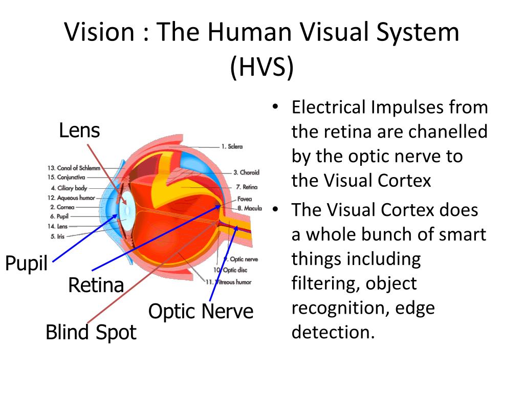
UAB School of Optometry Biology Diagrams The retina is the beginning of the nervous system's involvement with the visual system. It consists of five layers of neurons (described below). The pathway of information moves from the back of the eye towards the front, before exiting the back of the eye in an area called the optic disk. Visual information is passed directly through three
The tract splits into two. The larger visual fibers head to the lateral geniculate body. The smaller fibers for the light reflex pass between the lateral and medial geniculate bodies to the midbrain. Visual fibers. The lateral geniculate body of the posterior part of the thalamus contains the third-order neurons of the visual system. Of its 6

Basic Human Physiology Biology Diagrams
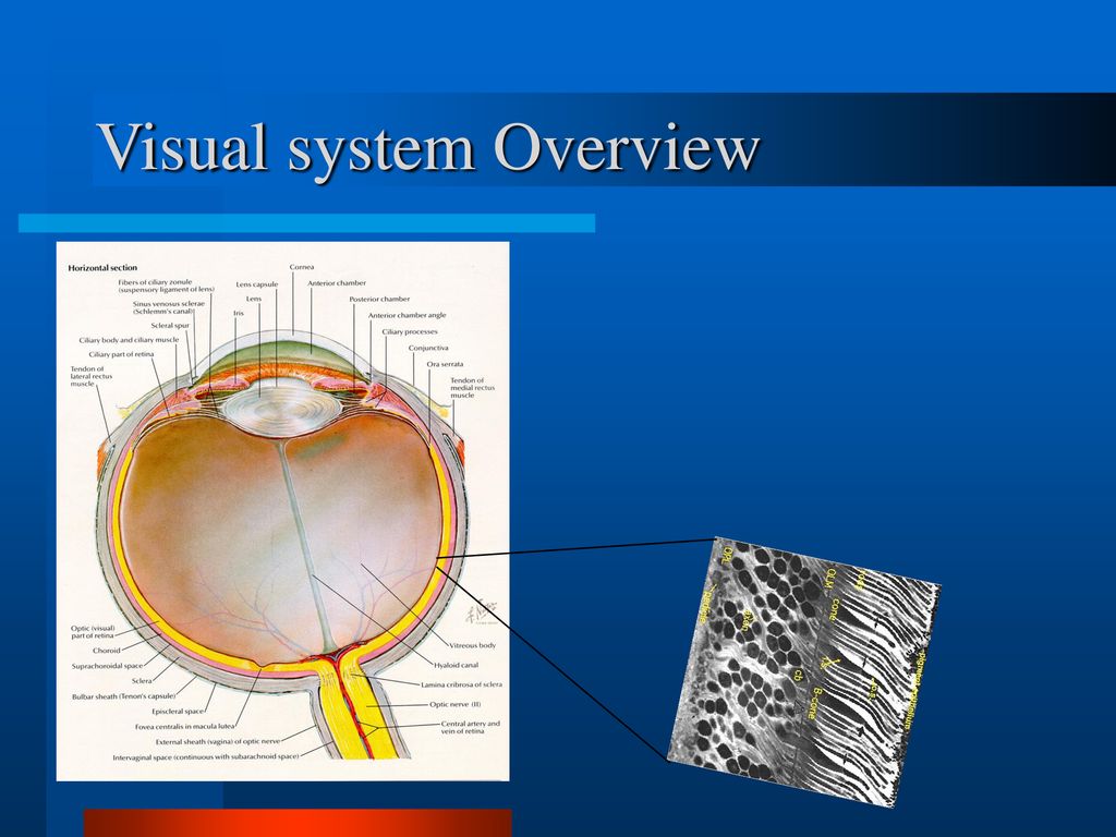
The visual system is the physiological basis of visual perception (the ability to detect and process light).The system detects, transduces and interprets information concerning light within the visible range to construct an image and build a mental model of the surrounding environment. The visual system is associated with the eye and functionally divided into the optical system (including

Learning Objectives. By the end of this section, you should be able to. 6.1.1 Describe the region of the electromagnetic spectrum that is perceived by our visual system, and the relative energy of photons at long and short wavelengths.; 6.1.2 Describe the major parts of the eye and their role in focusing light to create a clear image.; In this section, we will meet the range of the Both light and dark adaptation are crucial for maintaining visual function across a range of lighting conditions, demonstrating the remarkable adaptability of the human visual system. Adapted from Anatomy & Physiology by Lindsay M. Biga et al, shared under a Creative Commons Attribution-ShareAlike 4.0 International License, chapter 15. system, in combination with skilled examination, allows precise localization of neuropathological processes. Moreover, these principles guide effective diagnosis and management of neuro-ophthalmic disorders. The visual pathways perform the function of receiv-ing, relaying, and ultimately processing visual informa-tion.

Visual (Sight) System - Integrated Human Anatomy and ... Biology Diagrams
Aqueous humor is made by the ciliary body and flows along the posterior surface of the iris in the narrow posterior chamber. It then flows through the pupil and fills the anterior chamber. The scleral venous sinus (the old name is much more fun to say: the "canal of Schlemm") is a drainage system for the aqueous humor.